Research
Before acoustic models can be used clinically, for example to plan therapeutic ultrasound treatments, their accuracy must be tested against careful measurements. We have a programme of experimental validation that builds up step by step: from linear fields in water, through increasingly nonlinear fields, to propagation through tissue-mimicking and 3D-printed skull phantoms. Sources are characterised using acoustic holography and fed directly into k-Wave, and the predicted fields are compared with hydrophone measurements at the focus and beyond.
Alongside this, we study the measurements themselves: the repeatability and reproducibility of calibrated hydrophone measurements, the effect of hydrophone size and frequency response, and new sensors, such as the Fabry–Pérot ultrasound sensor developed by the UCL Photoacoustic Imaging Group, for mapping high-pressure focused fields beyond the range of conventional hydrophones. Data from this work are openly available through DRUM, our Data Repository for Ultrasound Metrology.
Low-intensity focused ultrasound can modulate neural activity non-invasively, with millimetre precision and the ability to reach deep brain targets through the intact skull. This makes it a promising tool both for neuroscience research and for the treatment of neurological and psychiatric disorders.
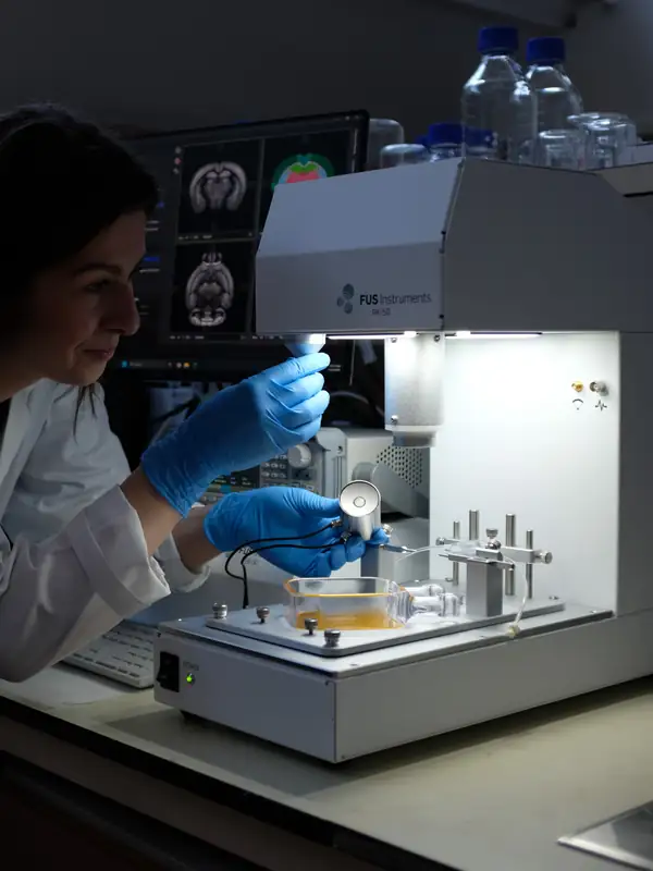
Preclinically, we run the UK’s first RK50 FUS system (FUS Instruments Inc.), a device for MRI-guided transcranial FUS in small animals. We use it to modulate brain activity, temporarily open the blood–brain barrier and deliver therapeutics to specific brain regions with millimetre accuracy, with the aim of developing surgery-free treatments for cancer, hypertension, and neurodegenerative and neuropsychiatric disorders.
We have also built a device for human neuromodulation in collaboration with the University of Oxford.
Agents such as microbubbles, nanodroplets and drug-loaded nanomedicines can respond strongly to ultrasound, so their effects can be switched on at a precise location in the body. They can be used to enhance image contrast, to open the blood–brain barrier temporarily, and to release drugs where they are needed.
One example is a class of biocompatible nanodroplets, smaller than a micron, that we have engineered to emit a detectable acoustic signal at a precise pressure threshold. Injected into the bloodstream, they give a real-time, in situ pressure readout inside the brain. Combined with measurements of how the skull attenuates and distorts ultrasound, this lets us plan and monitor treatments for each individual, which we are now applying to neuromodulation for hypertension in pregnancy (Lea-Banks et al. 2024).
We have developed computer models that accurately and efficiently predict how ultrasound waves travel through tissue. Our work spans the governing equations (including nonlinear propagation and power-law acoustic absorption), numerical methods – in particular Fourier pseudospectral methods, which are well suited to large-scale, high-frequency problems – and fast implementations on GPUs and supercomputers in collaboration with Jiri Jaros and the Supercomputing Technologies Research Group at Brno University of Technology.
Much of this work is released as open-source software, most notably the k-Wave toolbox and the differentiable simulator j-Wave (see Software). These models are used around the world for therapy planning, neuromodulation and blood–brain barrier opening, and as forward models for image reconstruction. We validate them against carefully characterised experimental measurements, and the associated data are available through our Data Repository.
Cryopreservation could allow tissues and organs to be stored for long periods, but rewarming them quickly and uniformly enough to avoid damage from ice formation and thermal stress is a major challenge. Ultrasound offers a way to deposit heat throughout a volume of tissue in a controlled way.
We develop methods for forming images from ultrasound measurements, with a focus on photoacoustic tomography and ultrasound computed tomography. In photoacoustic tomography, short laser pulses generate ultrasound waves in tissue that absorbs light, such as blood. Reconstructing where these waves came from gives high-resolution 3D images, typically of the vasculature as blood is a strong absorber. By using several optical wavelengths there is the potential to obtain information about the tissue’s constituents and even functional information. In ultrasound tomography we reconstruct quantitative maps of tissue properties such as sound speed, which can in turn be used to improve photoacoustic images.
We work closely with the UCL Photoacoustic Imaging Group, and have long term collaborations with the UCL Department of Computer Science, Centrum Wiskunde & Informatica (CWI), Amsterdam, and the University of Eastern Finland.